
-
Miami, Florida
-
Mon-Fri 8 am- 6pm (EST)
Patients from all over the US come to see Dr. Trapana due to his world class expertise in Spine
For decades, spinal surgery carried a daunting reputation: long midline incisions, aggressive stripping of the stabilizing paraspinal muscles, significant intraoperative blood loss, and prolonged, painful hospital stays. Traditional “open” spinal fusion often relied heavily on two-dimensional fluoroscopy (X-ray) and manual anatomical landmarking, introducing risks of hardware malposition, neurological injury, or adjacent tissue trauma.
The paradigm of spine surgery has shifted through the convergence of Robotic-Assisted Navigation and Minimally Invasive Spine Surgery (MISS). Instead of working through wide, open surgical corridors, modern spine surgeons use computer-assisted robotic guidance arms to plan and execute screw and cage placement with sub-millimeter precision through small keyhole portals.
In South Florida, Dr. Evan Trapana is a fellowship-trained orthopedic spine surgeon and Assistant Professor of Orthopedic Spine Surgery at the University of Miami Miller School of Medicine and UHealth Tower. Having completed his orthopedic surgery residency at the University of Miami/Jackson Memorial Hospital followed by advanced fellowship training in spine surgery at the University of California San Diego (UCSD) Medical Center, Dr. Trapana specializes in motion preservation, disc arthroplasty, and robotic-assisted minimally invasive reconstruction. By combining preoperative 3D planning with real-time robotic guidance, Dr. Trapana minimizes muscle disruption, mitigates complication risks, and helps patients achieve faster postoperative mobilization.
The primary driver of prolonged postoperative back pain and disability after traditional spinal fusion is not the bone work itself, but the collateral trauma inflicted on the multifidus and erector spinae paraspinal muscle envelope:
[TRADITIONAL OPEN SURGERY]
──► Long midline skin incision
──► Extensive subperiosteal stripping of paraspinal muscles off lamina & facets
──► Prolonged mechanical retraction ──► Muscle ischemia, denervation, and atrophy
──► Postoperative fusion disease / chronic paraspinal fibrosis
VS.
[ROBOTIC-ASSISTED MISS PROTOCOL]
──► Micro-incisions (1 to 2 cm) tailored to instrumentation trajectories
──► Muscle-splitting tubular dilation (splitting along natural muscle planes, not cutting)
──► Continuous vascular perfusion maintained to paraspinal soft tissues
──► Preservation of posterior tension band and supraspinous/interspinous ligaments
By preserving the paraspinal musculature and posterior ligamentous complex, patients avoid long-term muscular denervation, reducing the chronic postoperative soreness often associated with traditional open spinal fusions.
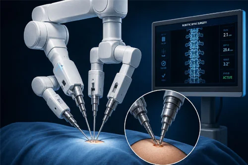
Robotic spine systems (such as the Globus ExcelsiusGPS® or Medtronic Mazor X™) do not operate autonomously. The surgeon maintains complete tactile and executive control. The robot functions as a high-precision mechanical guidance platform that translates a patient’s unique anatomy into sub-millimeter trajectory execution:
[PREOPERATIVE 3D VOLUMETRIC CT / INTRAOPERATIVE O-ARM SCAN]
│
▼
[PRE-SURGICAL SOFTWARE TRAJECTORY MAPPING]
(Surgeon selects exact screw diameter, length, and trajectory to avoid nerve roots)
│
▼
[DYNAMIC REAL-TIME OPTICAL REGISTRATION]
(Surveillance arrays track patient movement in real-time, matching coordinates)
│
▼
[ROBOTIC RIGID ARM DEPLOYMENT]
(Directs end-effector to the planned pedicle axis; sub-millimeter stability)
│
▼
[PERCUTANEOUS INSTRUMENTATION & INTERBODY FUSION]
(Cannulated drills, taps, and screws placed through muscle-sparing portals)
Pairing robotic stereotaxy with minimally invasive corridors yields marked clinical benefits:
┌──► 1. Sub-Millimeter Screw Accuracy (>98% to 99%)
│ (Eliminates pedicle breaches, nerve impingement, and CSF leaks)
│
[ROBOTIC MISS CLINICAL PROFILE] ──┼──► 2. Substantial Reduction in Radiation Exposure
│ (Replaces continuous live X-ray with computerized 3D mapping)
│
└──► 3. Accelerated Postoperative Mobilization
(Patients walking within 2 to 4 hours; outpatient or 24-hr discharge)
| Surgical Metric | Traditional Open Spine Surgery | Robotic-Assisted MISS Protocol |
| Incision Profile | 10 to 15+ cm single continuous incision | Multiple 1 to 2 cm percutaneous portals |
| Muscle Management | Stripped and detached off bone | Spared via muscle-splitting dilation |
| Screw Placement Accuracy | 88% to 94% (variable depending on visual landmarks) | > 98% to 99% sub-millimeter precision |
| Estimated Blood Loss (EBL) | 250 to 600+ mL (transfusion risks) | < 50 mL (minimal blood loss) |
| Hospital Length of Stay | 3 to 5 inpatient days | Outpatient / same-day or 24-hour stay |
| Narcotic Analgesic Dependency | 4 to 8+ weeks postoperatively | Minimal; rapid transition to non-opioid regimens |
| Return to Light Activity | 8 to 12 weeks | 2 to 4 weeks |
Robotic MISS is indicated for a broad spectrum of lumbar, thoracic, and cervical pathologies:
Spine surgery no longer necessitates long, debilitating recoveries. By utilizing robotic navigation platforms, 3D trajectory mapping, and muscle-sparing minimally invasive techniques, Dr. Evan Trapana provides patients with precise structural stabilization, decompression, and a streamlined path back to mobility and pain-free living.