
-
Miami, Florida
-
Mon-Fri 8 am- 6pm (EST)
Patients from all over the US come to see Dr. Trapana due to his world class expertise in Spine
Lumbar disc herniation is the leading cause of sciatica—a sharp, burning, or electric pain radiating from the lower back through the buttock, thigh, calf, and into the foot. While the majority of lumbar disc herniations resolve within six to eight weeks with non-operative care (physical therapy, anti-inflammatory medications, and epidural steroid injections), a significant subset of patients develop persistent, intractable pain or progressive motor deficits (such as foot drop). For these patients, mechanical compression of the traversing or exiting spinal nerve root must be addressed surgically.
Historically, traditional open discectomy required broad midline skin incisions, extensive detachment and stripping of the multifidus paraspinal muscle off the bony lamina, and resection of stabilizing spinal ligaments. This structural disruption often produced long-term muscular denervation, permanent tissue fibrosis, and persistent post-surgical back stiffness.
Today, Minimally Invasive Tubular Microdiscectomy (MISS Microdiscectomy) represents the gold standard for lumbar disc decompression. Utilizing muscle-splitting tubular dilation, high-definition microscopic visualization, and micro-instruments, spine surgeons can excise the herniated fragment through an operative corridor roughly the diameter of a dime.
In South Florida, Dr. Evan Trapana is a fellowship-trained orthopedic spine surgeon and Assistant Professor of Orthopaedic Spine Surgery at the University of Miami Miller School of Medicine and UHealth Tower. Dr. Trapana completed his orthopedic surgery residency at the University of Miami/Jackson Memorial Hospital followed by specialized fellowship training in spine surgery at the University of California San Diego (UCSD) Medical Center. Applying muscle-sparing tubular access and micro-decompressive techniques, Dr. Trapana delivers immediate nerve decompression while enabling patients to return home on the day of surgery with minimal post-procedural soreness.
Intervertebral discs act as hydraulic shock absorbers between the lumbar vertebrae. Each disc consists of a tough, fibrous outer ring (annulus fibrosus) encapsulating a gelatinous center (nucleus pulposus).
[Annular Tear / Fissure Formation]
│
▼
[Nucleus Pulposus Extrusion / Sequestration]
│
├─► 1. Mechanical Compression: Mass effect directly flattens nerve root
│
└─► 2. Chemical Radiculitis: Acidic disc glycoproteins trigger acute neuro-inflammation
│
▼
[Severe Sciatic Distribution Pain, Numbness, & Motor Weakness]
Sciatica involves dual mechanisms: mechanical distortion of the axon sheath and chemical radiculitis driven by inflammatory cytokines (TNF-$\alpha$, IL-6) released from the extruded nucleus pulposus.
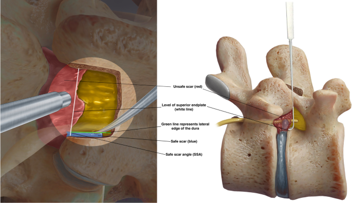
Minimally invasive tubular microdiscectomy avoids cutting or detaching paraspinal muscles. Instead, it uses serial dilation to separate muscle fibers along their natural planes:
[Flouro/Navigation Guidance: Localize Level via 15 mm Incision]
│
▼
[Serial Muscle Dilation (Multifidus fibers separated, not stripped)]
│
▼
[Tubular Retractor Docked onto Lamina / Interlaminar Window]
│
▼
[Operating Microscope / Endoscope Illumination (High 3D Magnification)]
│
▼
[Micro-Laminotomy & Ligamentum Flavum Flap Reflected]
│
▼
[Nerve Root Retraction ──► Disc Fragment Excision & Annular Inspection]
| Clinical Parameter | Traditional Open Discectomy | Tubular MISS Microdiscectomy |
| Incision Size | 3 to 6 cm midline incision | 1.4 to 1.8 cm keyhole portal |
| Muscle Trauma | Extensive subperiosteal strip & devascularization | Muscle-splitting dilation (Zero muscle cut) |
| Facet Joint Disruption | Increased risk of facet destabilization | Preserved; leaves posterior motion unit intact |
| Estimated Blood Loss | 50 to 150 mL | Minimal (< 10 to 20 mL) |
| Operative Setting | Inpatient stay (1 to 2 days) | True outpatient (Discharge in 2–3 hours) |
| Postoperative Narcotic Use | Moderate to heavy (1 to 3+ weeks) | Minimal; rapid transition to NSAIDs / Tylenol |
| Return to Light Work | 4 to 8 weeks | 1 to 2 weeks |
While elective microdiscectomy is pursued after conservative failure, immediate emergency decompression is required if a disc herniation causes Cauda Equina Syndrome (CES):
┌──► 1. Saddle Anesthesia (Numbness across perineum & inner thighs)
│
[EMERGENT SURGICAL RED FLAGS] ────┼──► 2. Bowel or Bladder Incontinence / Acute Urinary Retention
│
└──► 3. Rapidly Progressive Bilateral Motor Weakness (Flaccid paralysis)
Clinical Urgency: Cauda Equina Syndrome represents a surgical emergency. Decompression within 24 to 48 hours of onset is critical to avoid permanent bowel/bladder paralysis and irreversible sexual dysfunction.
Because tubular microdiscectomy bypasses muscle stripping, recovery is rapid:
Severe sciatica and debilitating disc herniations no longer require large open back surgeries, muscle detachment, or extended hospital stays. Through minimally invasive tubular microdiscectomy, Dr. Evan Trapana provides targeted neurological decompression through a small keyhole incision—delivering immediate sciatica relief while preserving spinal stability and accelerating return to everyday life.