
-
Miami, Florida
-
Mon-Fri 8 am- 6pm (EST)
Patients from all over the US come to see Dr. Trapana due to his world class expertise in Spine
Cervical radiculopathy—nerve root compression in the neck causing radiating arm pain, numbness, tingling, and motor weakness—is one of the most common spinal disorders in active adults. When conservative treatments such as physical therapy, anti-inflammatory medications, and targeted epidural steroid injections fail to relieve neural impingement, surgical decompression becomes the definitive pathway to prevent irreversible nerve injury.
For over half a century, the unchallenged benchmark for treating symptomatic single- or two-level cervical disc herniation was Anterior Cervical Discectomy and Fusion (ACDF). While ACDF reliably relieves nerve compression, eliminating motion across a spinal segment increases mechanical stress on the adjacent levels above and below.
To overcome this limitation, modern spine surgery developed Cervical Disc Arthroplasty (CDA), also known as total disc replacement (TDR). By replacing the damaged, herniated intervertebral disc with a mobile, engineered prosthetic joint rather than fusing it solid, CDA preserves natural cervical kinematics and reduces the risk of adjacent-segment degeneration.
In South Florida, Dr. Evan Trapana is a fellowship-trained orthopedic spine surgeon and Assistant Professor of Orthopaedic Spine Surgery at the University of Miami Miller School of Medicine and UHealth Tower. Having completed spine fellowship training at the University of California San Diego (UCSD) Medical Center, Dr. Trapana specializes in motion preservation, cervical disc arthroplasty, and minimally invasive spinal reconstruction. He evaluates each patient’s spinal alignment, bone quality, and facet joint integrity to guide them to the optimal intervention: dynamic motion preservation or durable fusion.
The cervical spine consists of seven vertebrae separated by shock-absorbing intervertebral discs. When an annulus fibrosus tears or degenerates, inner nucleus pulposus material herniates posterolaterally into the neuroforamen, compressing the exiting cervical spinal nerve root:
[Degenerated / Herniated Cervical Disc] ──► Mechanical Nerve Root Impingement (Neuroforamen)
│
▼
[Local Inflammatory Cascade] ──► Severe radiating arm pain (Radiculopathy)
│
▼
[Motor / Sensory Deficit] ──► Numbness in dermatomes & triceps/biceps weakness
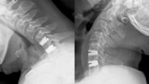
In an ACDF, the surgeon accesses the spine through a small anterior horizontal skin crease incision in the front of the neck:
[Anterior Micro-Incision] ──► Complete Discectomy & Foraminotomy (Decompresses Nerves)
│
▼
[Interbody Cage Placement] ──► Bone Graft / PEEK / Titanium Cage inserted into disc space
│
▼
[Anterior Plate & Screws] ──► Rigid fixation across the disc space ──► Bony Fusion
While ACDF provides predictable decompression, immobilizing a vertebral level alters the biomechanics of the neck. The fused level can no longer flex, extend, or rotate, forcing the adjacent discs above and below to absorb additional mechanical strain and intradiscal pressure. Long-term studies show that up to 25% of fusion patients develop symptomatic Adjacent Segment Disease (ASD) within 10 years, frequently requiring secondary operations.
Cervical Disc Arthroplasty removes the offending disc and replaces it with a dynamic, biocompatible artificial disc that mimics natural physiological movement:
┌──► 1. Bilateral Flexion and Extension
│
[CDA KINEMATIC FREEDOM RESTORED] ─┼──► 2. Unrestricted Lateral Bending
│
└──► 3. Axial Rotation (Torque Absorption)
[Anterior Micro-Incision] ──► Micro-Discectomy & Direct Nerve Decompression
│
▼
[Footprint Sizing & Keel Preparation] ──► Preserves native uncovertebral architecture
│
▼
[Artificial Disc Implantation] ──► Metal-on-Polymer (e.g., Mobi-C, Simplify, ProDisc-C)
│
▼
[Dynamic Motion Retained] ──► Normalizes load transfer across adjacent levels
| Clinical Parameter | Anterior Cervical Discectomy & Fusion (ACDF) | Cervical Disc Arthroplasty (CDA / TDR) |
| Surgical Goal | Neural decompression + permanent fusion | Neural decompression + motion preservation |
| Index Segment Motion | 0° (Completely immobilized) | Retains 6° to 12° physiological range of motion |
| Adjacent Segment Strain | Increased intradiscal pressure; higher wear | Normal physiologic load sharing; lower ASD risk |
| Hardware Failure Risk | Pseudarthrosis (non-union of bone graft) | No bone healing required; zero pseudarthrosis risk |
| Post-Operative Collar | Often required for 2 to 6 weeks | Rarely needed; immediate active range of motion |
| Return to Normal Activity | 6 to 12 weeks (waiting for bony fusion) | 3 to 6 weeks (earlier return to work and athletics) |
| Long-Term Reoperation Rate | Higher secondary surgery rates over 10 years | Significantly lower revision rate for adjacent wear |
While cervical disc arthroplasty provides distinct mechanical advantages, it is not universally applicable to all neck conditions. Patient selection is key:
Because CDA does not rely on biological bone graft consolidation (healing bone to bone), the recovery timeline is typically shorter than with a fusion:
Cervical radiculopathy and disc herniations no longer require permanent fusion of your neck. Through cervical disc arthroplasty, patients can achieve direct neural decompression and long-term symptom relief while preserving their natural range of motion and protecting adjacent spinal discs.