-
Miami, Florida
-
Mon-Fri 8 am- 6pm (EST)
Patients from all over the US come to see Dr. Trapana due to his world class expertise in Spine
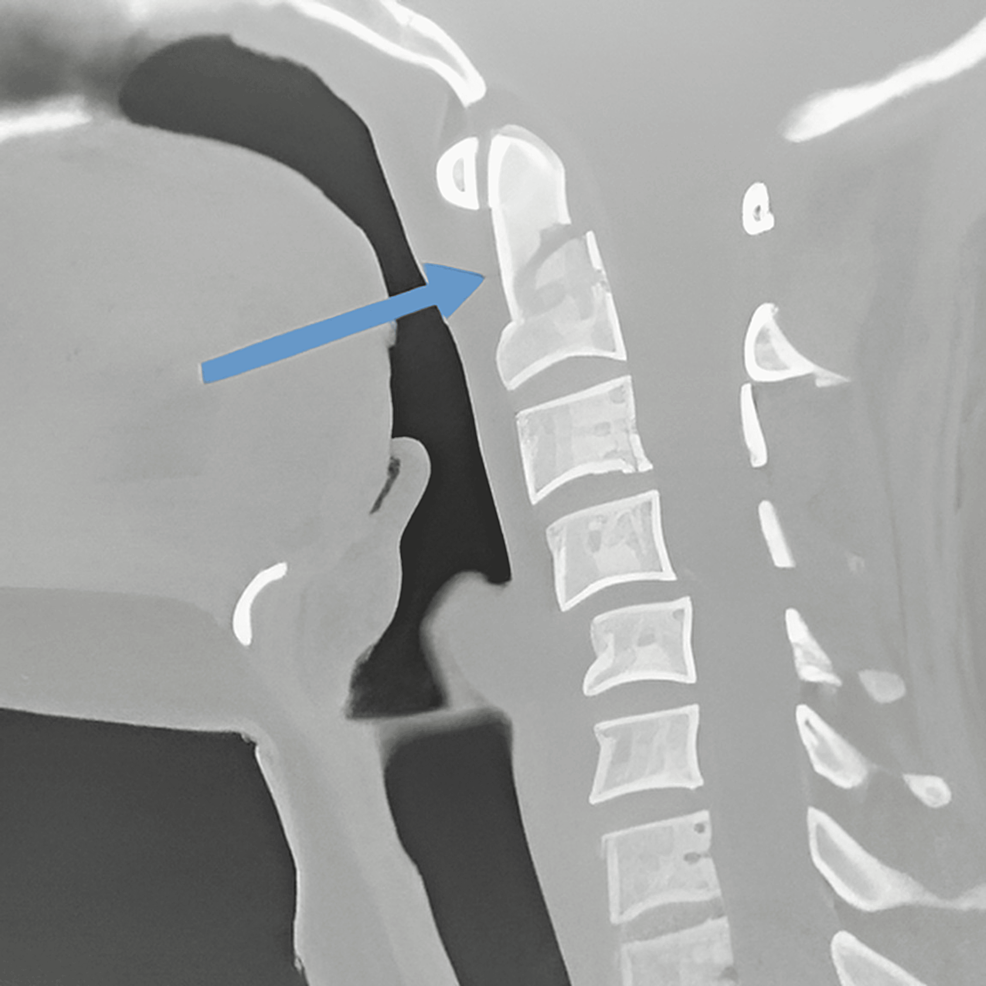
Fractures of the odontoid process (the dens of the second cervical vertebra, C2) represent the most frequent cervical spine injury in elderly adults and account for roughly 10% to 15% of all cervical spine fractures overall. The atlantoaxial complex (C1–C2) provides more than 50% of total cervical rotational mobility, but this exceptional range of motion relies on a delicate balance between bone geometry and ligamentous restraints rather than intervertebral disc buffers.
When traumatic forces shear the odontoid peg, mechanical stability is immediately compromised. In younger patients, these injuries typically result from high-energy blunt trauma, such as motor vehicle collisions or sports impacts. In older adults with osteopenia or osteoporosis, however, an odontoid fracture often follows a seemingly trivial low-energy event—such as a fall from standing height with neck hyperextension.
Management of odontoid fractures requires nuanced decision-making. Historically, non-operative management with rigid external immobilization (such as halo-vest orthoses) carried unacceptably high morbidity and mortality in elderly populations, while traditional posterior fusions permanently sacrificed half of the patient’s neck rotation.
In South Florida, Dr. Evan Trapana is a fellowship-trained orthopedic spine surgeon and Assistant Professor of Orthopaedic Spine Surgery at the University of Miami Miller School of Medicine and UHealth Tower. Having completed his orthopedic surgery residency at UM/Jackson Memorial Hospital and advanced spine fellowship training at the University of California San Diego (UCSD) Medical Center, Dr. Trapana specializes in upper cervical spine trauma, minimally invasive stabilization, and complex spinal reconstruction. Leveraging real-time 3D navigation, anterior motion-preserving screw fixation, and posterior atlantoaxial fusion protocols, Dr. Trapana tailors treatment to bone mineral quality, fracture orientation, and the patient’s physiological reserve.
The standard anatomical framework for evaluating odontoid fractures is the Anderson and D’Alonzo Classification, which stratifies injuries based on the anatomical level of the fracture line:
[ODONTOID FRACTURE CLASSIFICATION]
│
├─► Type I: Tip Avulsion (Rare, Alar ligament insertion)
│ └─► Generally stable; treated conservatively with a rigid collar
│
├─► Type II: Fracture at the Waist / Base of the Dens
│ └─► HIGH NON-UNION RATE (Up to 30%–85% with conservative care)
│
└─► Type III: Fracture Extends into C2 Vertebral Body Cancellous Bone
└─► High vascularity; heals reliably in a rigid cervical collar (85%–90% union)
▲ [Dens Tip] ────────► Type I: Alar Ligament Avulsion
/ \
| | [Waist / Neck] ───► Type II: High Non-Union / Watershed Zone
|___|
/ \
| C2 | [Body Base] ────► Type III: Cancellous Extension into Body
\_______/
Type II fractures occur through the narrow neck of the dens and present a persistent clinical challenge:
When a Type II fracture demonstrates instability, displacement $> 5\text{ mm}$, angulation $> 10^\circ$, or presents in an older adult where prolonged bed rest or external bracing is dangerous, surgical stabilization is warranted:
[DISPLACED TYPE II ODONTOID FRACTURE]
│
▼
[ASSESS TRANSVERSE LIGAMENT & MORPHOLOGY]
│
┌──────────────┴────────────────────────────────────────┐
▼ ▼
[INSPECT FRACTURE LINE & HABITUS] [ANTERIOR CONTRAINDICATIONS PRESENT]
│ │
├─► Fracture oblique: Anterior-superior ├─► Anterior-inferior oblique fracture
│ to posterior-inferior ├─► Chronic non-union (> 6 months)
├─► Intact transverse atlantal ligament ├─► Disrupted transverse atlantal ligament
├─► Non-osteoporotic bone purchase ├─► Severe barrel-chest / short neck anatomy
├─► No severe cervicothoracic kyphosis └─► Severe osteoporotic bone failure
│ │
▼ ▼
[ANTERIOR ODONTOID SCREW FIXATION] [POSTERIOR C1–C2 ARTHRODESIS]
(Preserves >50% Cervical Rotation) (Harms / Goel-C1 Lateral Mass + C2 Pedicle/Pars)
For acute, reducible fractures in suitable candidates, direct anterior screw osteosynthesis represents the definitive motion-preserving intervention:
[Supine Head-Extended Positioning under Dual Biplanar Fluoroscopy]
│
▼
[Anterior Retropharyngeal Approach via C5–C6 Transverse Neck Crease]
│
▼
[Working Corridor Medial to Carotid Sheath, Lateral to Trachea/Esophagus]
│
▼
[K-Wire Trajectory Drill across Anterior-Inferior C2 Body into Dens Tip]
│
▼
[Cannulated Herbert-Type Lag Screw Compresses Fracture Line]
When the fracture line is comminuted, chronicity exceeds several months, bone mineral density is poor, or the transverse atlantal ligament is torn (Dickman Type I injury), anterior screw fixation carries an unacceptably high risk of failure.
In these scenarios, Posterior C1–C2 Fusion using C1 Lateral Mass and C2 Pedicle/Pars Screws (Harms-Goel technique) provides rigid three-column biomechanical stabilization:
[Prone Rigid Mayfield Skull Pin Fixation]
│
▼
[Targeted Posterior Midline Suboccipital Exposure (C1 Arch & C2 Lamina)]
│
▼
[Navigation-Guided Screw Trajectory]
├─► C1: Polyaxial screws placed into bilateral C1 Lateral Masses
└─► C2: High-purchase screws into C2 Pedicles, Pars, or Translaminar corridors
│
▼
[Rigid Connecting Rods & Structural Autologous Bone Graft Placement]
| Clinical Feature | Conservative (Rigid Collar / Halo) | Anterior Odontoid Lag Screw | Posterior C1–C2 Fusion (Harms) |
| Cervical Rotation Preserved | Retained (if union occurs) | 100% retained (>50° bilateral rotation) | Sacrificed (~50% overall rotation lost) |
| Bony Union Rate | Low in Type II (30% to 60%) | High in acute cases (85% to 90%) | Exceptional (> 95% to 98%) |
| Halo-Vest Morbidity | High (Pin infection, pneumonia, dysphagia) | Eliminated | Eliminated |
| Surgical Incision | None | Small anterior neck crease (2.5–3 cm) | Midline suboccipital posterior incision |
| Primary Risk Profile | Pseudarthrosis, displacement, myelopathy | Screw pullout, loss of purchase in osteopenia | C2 nerve root irritation, loss of neck turning |
| Immediate Mobilization | Compromised by external brace | Rapid; light soft collar for comfort | Immediate; rigid internal screw stability |
The management of odontoid fractures in patients over the age of 65 requires special attention:
Upper cervical spine trauma and odontoid fractures demand nuanced management to balance bony union against long-term mobility. By offering both motion-preserving anterior odontoid screw fixation and rigid posterior atlantoaxial reconstruction, Dr. Evan Trapana tailors surgical care to the fracture morphology and the patient’s individual lifestyle and recovery goals.