
-
Miami, Florida
-
Mon-Fri 8 am- 6pm (EST)
Patients from all over the US come to see Dr. Trapana due to his world class expertise in Spine
Lumbar degenerative disc disease, adult degenerative scoliosis, and degenerative spondylolisthesis (slipped vertebrae) frequently converge on a common painful pathology: disc collapse with secondary central and neuroforaminal stenosis. Historically, achieving interbody fusion and nerve root decompression required posterior approaches, such as Posterior Lumbar Interbody Fusion (PLIF) or Transforaminal Lumbar Interbody Fusion (TLIF).
While effective, traditional posterior fusion techniques demand operating through the dorsal back muscles, retracting the thecal sac and exiting nerve roots, and dissecting near the epidural venous plexus. This posterior dissection can lead to paraspinal muscle denervation, perineural fibrosis, and prolonged postoperative back soreness.
Lateral Lumbar Interbody Fusion (LLIF)—also known by trade names such as Extreme Lateral Interbody Fusion (XLIF) or Direct Lateral Interbody Fusion (DLIF)—fundamentally changed this paradigm. By accessing the lumbar spine through a retroperitoneal corridor from the patient’s side (flank), surgeons can insert large interbody reconstruction cages that span the dense apophyseal ring of the vertebral body. This simultaneously restores disc height and straightens coronal deformity while achieving indirect neural decompression—expanding the pinched spinal canal and neuroforamina without directly exposing or retracting the delicate nerve roots.
In South Florida, Dr. Evan Trapana is a fellowship-trained orthopedic spine surgeon and Assistant Professor of Orthopaedic Spine Surgery at the University of Miami Miller School of Medicine and UHealth Tower. Having completed his orthopedic residency at UM/Jackson Memorial Hospital and advanced spine surgery fellowship training at the University of California San Diego (UCSD) Medical Center, Dr. Trapana specializes in robotic guidance, complex spinal deformity correction, and minimally invasive lateral retroperitoneal approaches. Through meticulous intraoperative neuromonitoring and lateral access surgery, Dr. Trapana reconstructs spinal stability and decompresses nerves while sparing posterior paraspinal musculature.
Traditional spine surgery relies on “direct” decompression: using rongeurs, drills, and curettes to mechanically carve away the bone spurs (osteophytes), lamina, and hypertrophied ligamentum flavum compressing the nerves.
In contrast, LLIF utilizes the principles of ligamentotaxis and structural disc restoration to achieve indirect decompression:
[DEGENERATIVE DISC COLLAPSE & STENOSIS]
(Loss of intervertebral height, bulging annulus, buckled ligamentum flavum, pinched foramen)
│
▼
[LATERAL INSERTION OF WIDE INTERBODY CAGE]
(Spans dense cortical apophyseal ring bilaterally across disc space)
│
├─► 1. Restores Intervertebral Disc Height
│ └─► Tenses annulus fibrosus & unbuckles ligamentum flavum
│
├─► 2. Expands Neuroforaminal Area by 30% to 50%
│ └─► Frees exiting nerve roots without entering the foramen
│
└─► 3. Reconstitutes Central Canal Dimensions
└─► Relieves thecal sac compression via natural ligamentotaxis
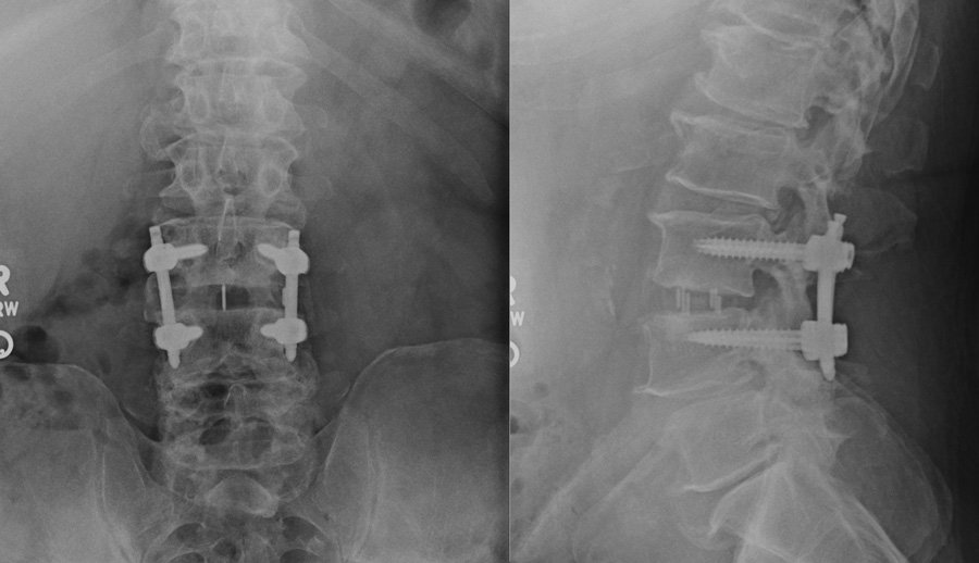
Executing an LLIF requires navigating through the retroperitoneal space and carefully transversing the psoas major muscle, which houses the sensitive nerves of the lumbosacral plexus:
[Lateral Decubitus Positioning & True AP/Lateral Fluoroscopy]
│
▼
[Flank Micro-Incision (3 to 5 cm) & Retroperitoneal Finger Dissection]
│
▼
[Docking on Psoas Major Muscle with Real-Time Directional EMG]
│
▼
[Muscle-Splitting Dilation Avoiding Lumbosacral Plexus]
│
▼
[Wide Discectomy, Endplate Prep, & Interbody Graft Delivery]
│
▼
[Percutaneous Robotic Posterior Pedicle Screw Fixation]
While lateral surgery provides exceptional access across the mid- and upper-lumbar spine (L1–L2 down through L4–L5), it has a clear anatomical boundary at the L5–S1 junction:
┌──► 1. Iliac Crest Blockade: High iliac wings block a direct lateral trajectory
│
[WHY L5–S1 CANNOT BE ACCESSED] ───┼──► 2. Great Vessel Bifurcation: Vena cava and aorta divide into iliac vessels
│
└──► 3. Lumbosacral Trunk: Major lower-extremity motor roots drape over disc
The Clinical Workaround: When fusion is required down to the sacrum (L5–S1), Dr. Trapana frequently combines an Anterior Lumbar Interbody Fusion (ALIF) or a minimally invasive Transforaminal Lumbar Interbody Fusion (MIS-TLIF) at L5–S1 with an LLIF at the levels above (L2–L5), creating a unified reconstructive construct.
| Surgical Metric | Transforaminal Fusion (TLIF/PLIF) | Lateral Interbody Fusion (LLIF/XLIF) |
| Corridor of Access | Posterior midline through back muscles | Lateral retroperitoneal flank corridor |
| Paraspinal Muscle Trauma | Muscle stripped, retracted, or dilated | Zero posterior back muscle disruption |
| Decompression Type | Direct (laminectomy, facetectomy, flavectomy) | Indirect (disc height restoration & ligamentotaxis) |
| Interbody Cage Size | Small (8–10 mm wide; sits centrally) | Massive (18–26 mm wide; spans cortical ring) |
| Subsidence Risk | Moderate to high (rests on softer bone) | Very low (anchored to dense apophyseal ring) |
| Deformity / Scoliosis Correction | Limited coronal restoration per level | Superior coronal & lordotic realignment |
| Thecal Sac / Dura Exposure | Direct exposure (CSF leak risk) | Zero dural exposure; no epidural scar tissue |
Lateral lumbar interbody fusion is indicated for conditions characterized by mechanical instability, severe disc space collapse, and deformity:
Because LLIF bypasses the major posterior muscle groups of the back, patients avoid the deep muscular soreness typical of traditional posterior fusions:
Lateral Lumbar Interbody Fusion (LLIF/XLIF) represents a major advancement in minimally invasive spine reconstruction. By restoring disc height, untethering compressed nerves through natural ligamentotaxis, and sparing the muscles of the lower back, Dr. Evan Trapana offers patients durable spinal stability and decompression with reduced surgical trauma.