
-
Miami, Florida
-
Mon-Fri 8 am- 6pm (EST)
Patients from all over the US come to see Dr. Trapana due to his world class expertise in Spine
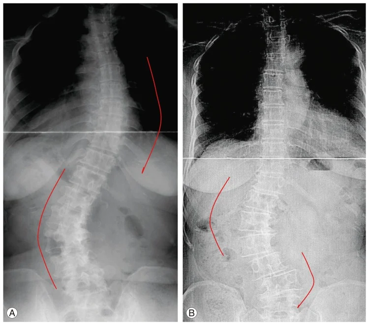
Unlike adolescent idiopathic scoliosis, which develops primarily during the pubertal growth spurt in an otherwise healthy skeleton, Adult Degenerative Scoliosis (ADS)—also termed de novo scoliosis—arises later in life as a consequence of asymmetric disc degeneration, facet joint arthrosis, and progressive osteoporosis. As intervertebral discs collapse unevenly, the structural foundation of the lumbar spine destabilizes, producing rotatory subluxation, lateral listhesis (slipping), and coronal curvature.
However, modern spinal deformity research has confirmed a critical clinical truth: the lateral (coronal) curve is rarely the primary driver of disability. Instead, patient-reported pain, chronic fatigue, and functional limitations correlate directly with the loss of sagittal balance—the forward pitch of the trunk that occurs when natural lumbar lordosis collapses. Patients find themselves expending immense muscular energy simply to maintain an upright horizontal gaze, leading to chronic paraspinal muscle exhaustion, severe neurogenic claudication, and progressive loss of independent ambulation.
Historically, correcting complex adult spinal deformities required extensive, open, multi-level osteotomies through broad posterior incisions, which carried high blood loss, substantial infection rates, and high pseudarthrosis risk. Today, advanced spinopelvic parameter alignment, computer-guided robotic navigation, and hybrid minimally invasive surgical (cMIS) techniques have redefined deformity correction.
In South Florida, Dr. Evan Trapana is a fellowship-trained orthopedic spine surgeon and Assistant Professor of Orthopaedic Spine Surgery at the University of Miami Miller School of Medicine and UHealth Tower. Having completed his orthopedic surgery residency at the University of Miami/Jackson Memorial Hospital and advanced spine surgery fellowship training at the University of California San Diego (UCSD) Medical Center, Dr. Trapana specializes in complex spinal deformity, robotic reconstruction, and motion-sparing realignments. Through precise spinopelvic planning and tailored corrective osteotomies, Dr. Trapana reconstructs global spinal balance to relieve mechanical pain, decompress nerve roots, and restore upright posture.
Evaluating adult spinal deformity begins not with localized lumbar X-rays, but with full-length, 36-inch standing radiographs capturing the entire skeletal axis from the skull to the femoral heads. Spinal balance is governed by a tightly coupled geometric relationship between the spine and the pelvis:
┌──► 1. Pelvic Incidence (PI)
│ ──► Fixed anatomical angle of pelvic morphology (does not change)
│
[SPINOPELVIC BALANCE PARAMETERS] ─┼──► 2. Pelvic Tilt (PT)
│ ──► Compensatory retroversion/rotation of the pelvis (Target < 20°)
│
└──► 3. Lumbar Lordosis (LL)
──► Inward inward curve matching the pelvis: Target LL = PI ± 10°
[DEGENERATIVE DISC & FACET COLLAPSE]
│
▼
[LOSS OF LUMBAR LORDOSIS (Flatback Deformity)]
│
▼
[SAGITTAL VERTICAL AXIS (SVA) SHIFTS FORWARD (> 5 cm)]
(The C7 plumb line falls far in front of the posterior superior sacral margin)
│
▼
[COMPENSATORY FATIGUE MECHANISMS]
(Pelvic retroversion [High PT], knee flexion, thoracic hypokyphosis ──► Severe Muscular Exhaustion)
Adult degenerative scoliosis is rarely a purely cosmetic or structural issue—it is almost always complicated by debilitating neurological compression:
[ASYMMETRIC DISC COLLAPSE & ROTATORY SUBLUXATION]
│
┌────────────────┴────────────────┐
▼ ▼
[CONCAVE APEX COMPRESSION] [CONVEX TRACTION RADICULOPATHY]
(Bone spurs & asymmetric disc (Exiting nerve roots stretched over
telescope into foramen ──► the structural apex of the curve)
Severe foraminal stenosis)
Rather than relying solely on high-morbidity open posterior approaches, Dr. Trapana frequently utilizes circumferential minimally invasive surgery (cMIS) or hybrid reconstructive protocols to minimize surgical trauma while achieving global alignment:
[STAGE 1: LATERAL ANTERIOR RECONSTRUCTION (LLIF / ALIF)]
(Multi-level lateral disc access spares posterior muscle, releases rigid curves, & restores lordosis)
│
▼
[STAGE 2: PERCUTANEOUS ROBOTIC POSTERIOR FIXATION]
(Sub-millimeter pedicle screw navigation across deformity curves through keyhole portals)
│
▼
[STAGE 3: TARGETED POSTERIOR OSTEOTOMIES (As Needed for Rigid Curves)]
(Schwab Grade 1/2 facetectomies to close posterior columns and lock in sagittal balance)
Through small retroperitoneal flank incisions, large lordotic interbody spacers are placed across the collapsed apex levels.
In stiff, fixed adult deformities, releasing anterior discs alone may not provide adequate lordosis. Through targeted posterior releases, the facet joints, ligamentum flavum, and spinous processes are excised across multiple levels (Schwab Grade 1 and 2 osteotomies). When the spine is subsequently extended and secured to contoured rods, the posterior gaps close, producing 5 to 10 degrees of additional segmental lordosis per level.
Scoliotic anatomy features dysplastic, rotated, and hypoplastic pedicles that challenge conventional freehand instrumentation. Dr. Trapana employs real-time 3D navigation and robotic stereotaxy to guide pedicle screws through small portals with sub-millimeter precision, protecting neurological structures and ensuring stable anchor purchase across poor-quality osteoporotic bone.
| Surgical Metric | Traditional Full-Open Correction | Modern Hybrid / cMIS Protocol |
| Surgical Exposure | Extensive midline incision (T10 to Pelvis) | Targeted lateral incisions + percutaneous ports |
| Paraspinal Muscle Detachment | Complete bilateral muscle stripping/ischemia | Spared via retroperitoneal and muscle-splitting portals |
| Estimated Blood Loss (EBL) | 1,500 to 3,000+ mL (frequent transfusions) | < 300 to 500 mL (substantially lower transfusion rates) |
| Indirect Decompression | Rare; relies on multi-level open laminectomies | Primary; wide cages decompress foramen via ligamentotaxis |
| ICU & Hospital Stay | 7 to 10+ days (extended ICU recovery) | 3 to 5 days (often bypassing the ICU entirely) |
| Hardware Accuracy | 88% to 92% (in complex rotatory curves) | > 98% with real-time 3D robotic navigation |
| Pseudarthrosis Risk | Higher risk in long posterior-only constructs | Lower; massive surface area across wide lateral cages |
When adult deformity corrections extend down into the lower lumbar spine, ending a fusion construct at L5 carries an unacceptably high rate of mechanical failure, pseudarthrosis, and subsequent L5–S1 disc collapse.
Adult Degenerative Scoliosis and loss of sagittal balance are complex structural disorders that do not have to result in permanent spinal hunching, debilitating claudication, or chronic pain. Through modern spinopelvic parameter alignment, minimally invasive lateral releases, and robotic-assisted navigation, Dr. Evan Trapana provides patients with comprehensive reconstructive solutions designed to restore global balance, decompress nerves, and return patients to an active, upright life.