
-
Miami, Florida
-
Mon-Fri 8 am- 6pm (EST)
Patients from all over the US come to see Dr. Trapana due to his world class expertise in Spine
Cervical Spondylotic Myelopathy (CSM) is the most common cause of non-traumatic spinal cord dysfunction in adults worldwide. Unlike cervical radiculopathy—which involves the compression of an isolated peripheral nerve root causing sharp, radiating pain down an arm—myelopathy is a disorder of the central spinal cord itself. It is insidious, progressive, and frequently misdiagnosed in its early stages as normal aging, carpal tunnel syndrome, peripheral neuropathy, or general joint stiffness.
As the cervical spine undergoes age-related degeneration, bulging intervertebral discs, hypertrophied ligamentum flavum, and expanding osteophytic bone spurs gradually narrow the spinal canal. When mechanical narrowing reaches a critical threshold, it exerts direct chronic pressure on the spinal cord and compromises its microvascular blood supply. Left untreated, prolonged cord compression causes permanent gray and white matter ischemia, neuronal cell death, and irreversible spinal cord atrophy (myelomalacia).
In South Florida, Dr. Evan Trapana is a fellowship-trained orthopedic spine surgeon and Assistant Professor of Orthopaedic Spine Surgery at the University of Miami Miller School of Medicine and UHealth Tower. Having completed his orthopedic surgery residency at the University of Miami/Jackson Memorial Hospital followed by specialized spine surgery fellowship training at the University of California San Diego (UCSD) Medical Center, Dr. Trapana specializes in advanced cervical reconstruction and decompression. By combining high-resolution MRI diagnostic mapping with motion-sparing and minimally invasive decompressive approaches, Dr. Trapana aims to halt neurological decline and restore patient function before permanent cord atrophy sets in.
The progression of cervical spondylotic myelopathy involves both static anatomical constraints and dynamic, motion-induced trauma:
[DEGENERATIVE CERVICAL SPONDYLOSIS]
(Disc collapse, osteophyte formation, ligamentum flavum buckling)
│
▼
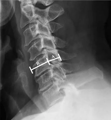
[STATIC SPINAL CANAL STENOSIS (< 10–12 mm AP Diameter)]
│
▼
[DYNAMIC REPETITIVE CORD CONTUSION]
(Neck flexion stretches cord over osteophytes; extension pinches cord against buckled ligament)
│
▼
[MICROVASCULAR ISCHEMIA & ENDOTHELIAL FAILURE]
(Compression of anterior spinal artery & sulco-commissural microvessels)
│
▼
[PERMANENT MYELOMALACIA & GLIAL SCARRING (Spinal Cord Atrophy)]
Because CSM often develops without neck pain, patients rarely recognize the severity of their condition until significant cord function is compromised. Diagnosis relies on identifying subtle “long-tract” signs:
┌──► 1. Loss of Fine Motor Hand Coordination
│ (Difficulty buttoning shirts, tying shoes, handwriting deterioration)
│
[CSM DIAGNOSTIC HALLMARKS] ───────┼──► 2. Balance Instability and “Spastic” Gait
│ (Feeling unsteady in the dark, heavy/stiff legs, wide-based walking)
│
└──► 3. Pathological Upper Motor Neuron (UMN) Reflexes
(Hyperreflexia, positive Hoffmann sign, inverted radial reflex, clonus)
For conditions like acute lumbar sciatica or mild cervical radiculopathy, conservative treatments (physical therapy, chiropractic adjustments, oral steroids) are frequently effective first-line treatments.
For moderate-to-severe Cervical Spondylotic Myelopathy, conservative therapy is fundamentally ineffective. Physical therapy cannot expand a narrow bony spinal canal, and aggressive neck manipulation poses severe risks of acute cord injury.
Natural history studies demonstrate that CSM typically follows a stepwise, progressive neurological decline:
Dr. Evan Trapana personalizes the surgical corridor based on sagittal alignment, the number of compressive levels, and the exact location of the offending pathology (anterior vs. posterior):
[CERVICAL SPONDYLOTIC MYELOMALACIA CONFIRMED]
│
▼
[ASSESS CERVICAL SAGITTAL ALIGNMENT]
│
┌─────────────┴─────────────────────────────┐
▼ ▼
[LORDOTIC ALIGNMENT] [KYPHOTIC ALIGNMENT]
│ │
├─► 1–2 Levels: Anterior (ACDF / CDA) └─► ANTERIOR APPROACH MANDATORY
│ (Posterior decompression fails because cord
└─► 3+ Levels: Posterior remains tethered against anterior spurs)
(Laminoplasty vs. Laminectomy & Fusion) ──► Multi-level ACDF or Corpectomy (ACCF)
Best suited when compression is localized across 1 to 3 levels, or when the patient exhibits cervical kyphosis (forward curvature):
Ideal when compression spans multiple levels (3 or more cervical segments) in a patient with preserved lordosis:
| Surgical Approach | Primary Anatomical Indications | Motion Preservation | Lordosis Restoration | Risk Profile |
| Multilevel ACDF / ACCF (Anterior) | Kyphotic cervical spine; 1–3 levels of anterior compression | No (Solid segmental fusion) | Excellent (Direct structural lordosis reconstruction) | Transient dysphagia (swallowing difficulty) |
| Cervical Laminoplasty (Posterior) | Lordotic cervical spine; 3+ levels of multi-segmental stenosis | Yes (Preserves ~75%–85% of baseline motion) | Neutral (Relies on pre-existing natural lordosis) | Axial neck stiffness; C5 nerve root palsy (< 5%) |
| Laminectomy & Fusion (Posterior) | Multi-level stenosis with segmental instability or spondylolisthesis | No (Rigid screw-rod fixation) | Moderate (Dependent on surgical positioning) | Hardware strain; loss of neck rotation/flexion |
Spinal cord recovery is a gradual biological process. Following decompressive surgery:
Cervical Spondylotic Myelopathy is a progressive spinal condition that demands prompt clinical recognition and timely intervention. Waiting for severe balance loss, drop attacks, or irreversible hand clumsiness often means operating on an atrophied spinal cord. Through modern anterior reconstruction and posterior motion-preserving laminoplasty, Dr. Evan Trapana provides patients with decompressive pathways designed to safeguard neurological function and maintain independence.