
-
Miami, Florida
-
Mon-Fri 8 am- 6pm (EST)
Patients from all over the US come to see Dr. Trapana due to his world class expertise in Spine
Chronic lower back pain is one of the most common reasons adults seek medical care, yet identifying its exact anatomical root remains a clinical challenge. While herniated discs, spinal stenosis, and facet joint arthritis frequently dominate lumbar diagnostics, up to 30% of chronic lower back pain cases actually originate lower in the pelvis at the Sacroiliac (SI) Joint.
Because the symptoms of SI joint dysfunction mimic lumbar disc herniations and sciatica, many patients undergo years of ineffective treatments—or even unnecessary lumbar spine surgeries—without finding relief.
At the University of Miami Health System and Jackson Memorial Hospital, orthopedic spine specialist Dr. Evan Trapana utilizes targeted diagnostic protocols to differentiate true lumbar spine pathology from pelvic instability. By combining clinical provocative testing with image-guided diagnostic blocks and minimally invasive SI joint fusion, Dr. Trapana helps patients across South Florida resolve debilitating pelvic pain at its source.
The sacroiliac joints sit on either side of the lower spine, connecting the triangular sacrum at the base of the vertebral column to the ilium (pelvic bone).
[Lumbar Vertebrae] ──► Transfers Upper Body Weight
│
▼
[Sacroiliac Joints] ──► Shock-Absorbing Bridge (Transfers Load to Pelvis & Legs)
│
▼
[SI Joint Dysfunction] ──► Hypermobility (Instability) OR Hypomobility (Arthritis)
Unlike the lumbar spine, which is designed for significant bending and twisting, the SI joint is a weight-bearing, shock-absorbing structure designed for stability, allowing only 2 to 4 millimeters of micro-motion. SI joint dysfunction typically arises from two mechanical issues:
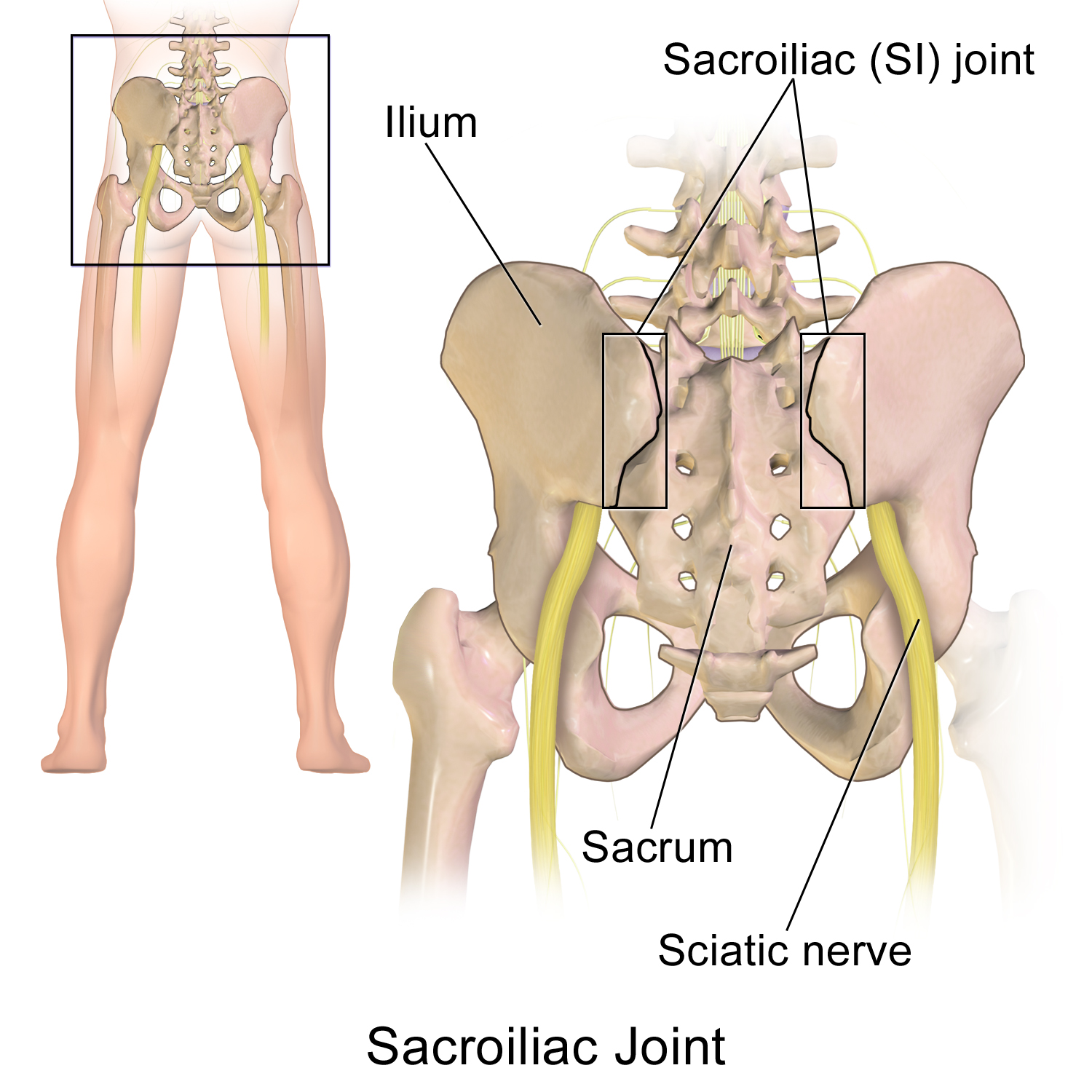
Because lumbar nerves exit directly above the pelvis, pain location alone can be misleading. Distinguishing between lumbar spine pain and SI joint dysfunction relies on specific clinical symptom patterns:
┌──► 1. Pain Located Below the L5 Level (Fortin Finger Point Test)
├──► 2. Unilateral Buttock / Groin Radiation (Rarely extends below knee)
[SI JOINT SYMPTOM PROFILE]─┼──► 3. Pain Exacerbated by Transitional Movements (Sitting to Standing)
├──► 4. Asymmetric Single-Leg Stance Sensitivity (Stairs / Car Entry)
└──► 5. Absence of True Neurological Deficits (Normal reflexes/strength)
When asked to pinpoint their worst pain, patients with SI joint dysfunction consistently point to a region directly over the posterior superior iliac spine (PSIS)—a spot just below the beltline—rather than the central lumbar spine.
SI joint pain is classically triggered by transitional movements that stress the pelvis, such as standing up from a deep chair, getting out of a car, climbing stairs, or rolling over in bed.
While lumbar sciatica typically shoots down the back of the leg past the knee and into the foot, SI joint pain radiates into the upper buttock, groin, or upper thigh, rarely extending past the knee joint.
Standard X-rays and MRI scans frequently fail to show SI joint pain because mechanical instability is a dynamic issue rather than a static anatomical deformity. Dr. Trapana uses a two-stage diagnostic framework:
A cluster of specialized physical manipulation maneuvers (including FABER, Gaenslen’s, Compression, Distraction, and Thigh Thrust tests) stresses the SI joint. Reproducing the patient’s familiar pain on three or more of these tests strongly indicates an SI joint origin.
To confirm the diagnosis, an image-guided (fluoroscopic or ultrasound) injection delivers a small dose of local anesthetic directly into the joint space. If the injection provides immediate, significant pain relief while the anesthetic is active, it confirms the SI joint as the primary pain generator.
| Clinical Factor | Lumbar Disc / Stenosis Pathology | Sacroiliac (SI) Joint Dysfunction |
| Primary Pain Center | Central lower back or L4–S1 region | Below beltline (buttock / PSIS area) |
| Leg Radiation | Shoots down back of leg past the knee | Radiates into buttock, hip, or groin |
| Neurological Deficit | Possible foot drop, numbness, or reflex loss | Zero neurological motor deficits |
| Pain Trigger | Forward bending or prolonged sitting | Transitional movements (chair to standing) |
| Diagnostic Gold Standard | Diagnostic MRI showing nerve compression | Fluoroscopic diagnostic joint injection |
When conservative management (physical therapy, pelvic stabilization belts, and targeted anti-inflammatory injections) fails to provide lasting relief, surgical stabilization offers a definitive solution:
Persistent lower back and buttock pain that fails to respond to standard spine treatments requires a fresh diagnostic look. By pinpointing SI joint instability through clinical testing and precision injections, Dr. Evan Trapana offers patients across Miami and South Florida a clear path to lasting pelvic stability and pain-free mobility.