
-
Miami, Florida
-
Mon-Fri 8 am- 6pm (EST)
Patients from all over the US come to see Dr. Trapana due to his world class expertise in Spine
When a lumbar vertebra slips forward out of its natural alignment over the bone beneath it, the resulting condition is known as spondylolisthesis. Whether caused by age-related joint degeneration (degenerative spondylolisthesis) or a structural stress fracture in the pars interarticularis (isthmic spondylolisthesis), this forward displacement narrows the spinal canal and nerve exits. Patients often experience a combination of severe lower back pain and neurogenic claudication—cramping, numbness, or aching in the legs that worsens when walking or standing upright.
For decades, the standard surgical dogma dictates that treating spondylolisthesis requires a two-part approach: decompressing the pinched nerves and performing a rigid spinal fusion with screws, rods, and bone grafts to prevent the slippage from progressing.
However, modern advancements in biomechanics and ultra-minimally invasive techniques are challenging the notion that every slipped vertebra demands fusion. At the University of Miami Health System and Jackson Memorial Hospital, orthopedic spine surgeon Dr. Evan Trapana utilizes careful stability criteria to determine when motion-preserving standalone decompression is safe—and when advanced, minimally invasive robotic fusion is required.
Selecting the right surgical option depends heavily on understanding the underlying structural mechanism driving the slippage:
[Degenerative Spondylolisthesis] ──► Facet Joint & Disc Breakdown ──► Often “Stable” Slip
VS.
[Isthmic Spondylolisthesis] ──► Pars Fracture / Bony Defect ──► Inherently “Unstable” Slip
To answer whether fusion is necessary, Dr. Trapana evaluates the dynamic stability of the affected spinal segment.
[Dynamic Flexion / Extension X-Rays]
│
├─► < 3–4 mm Motion (Stable) ──► Standalone Motion-Preserving Decompression
│
└─► > 4 mm Motion or Angular Tilt (Unstable) ──► Minimally Invasive Robotic Fusion
If standing flexion/extension radiographs show that the slipped vertebra remains rigid without abnormal movement, the spine is considered mechanically stable. In these selective cases, forcing a rigid fusion may be unnecessary.
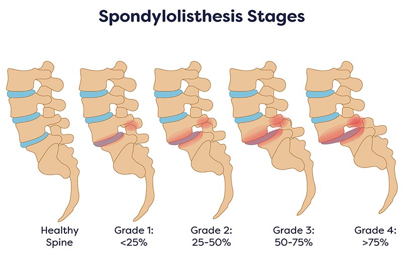
For stable, low-grade degenerative slips, Dr. Trapana utilizes muscle-sparing endoscopic or tubular micro-laminotomies. By removing only the specific hypertrophic ligament and bone spurs pinching the nerves while leaving the central midline structures and facet joints intact, the nerve pain is relieved while natural back flexibility is preserved.
When flexion/extension views reveal gross mechanical instability (greater than 3 to 4 millimeters of active movement), high-grade slippage, or severe axial mechanical back pain, stabilization is essential to prevent secondary nerve injury. In these scenarios, Dr. Trapana deploys 3D navigation and robotic guidance to place pedicle screws and interbody cages through micro-incisions, restoring alignment while minimizing soft-tissue disruption.
| Clinical Feature | Standalone Motion-Preserving Decompression | Minimally Invasive / Robotic Spinal Fusion |
| Primary Indication | Stable slip (< 3 mm motion); primary leg pain | Unstable slip (> 4 mm motion); severe back pain |
| Hardware Used | Zero hardware (screws or rods) | Precision pedicle screws & interbody cages |
| Back Mobility | 100% natural motion preserved at segment | Movement eliminated at treated level to fix slip |
| Adjacent Segment Stress | Low risk of accelerated wear above/below | Higher mechanical load transferred to neighboring discs |
| Surgical Incisions | Single micro-incision (< 1 cm) | Few micro-incisions (1–2 cm each) |
| Recovery & Discharge | Same-day outpatient discharge (1–2 hours) | Short hospital stay or same-day discharge |
A diagnosis of spondylolisthesis does not automatically mean you need metal screws, rods, or a fused back. By utilizing dynamic imaging to differentiate between stable and unstable spinal slips, Dr. Evan Trapana provides patients across Miami and South Florida with personalized, motion-conscious care designed to restore nerve function and maintain quality of life.