
• Miami, Florida

  • Mon-Fri 8 am- 6pm (EST)

Endoscopic Lumbar Foraminotomy: Targeted Relief for Lateral Recess Stenosis and Pinched Exit Nerves

Severe radiating leg pain, burning sensations, or numbness that worsens when walking or standing is often rooted in a very specific anatomical bottleneck: neural foraminal and lateral recess stenosis. The neural foramen is the small bony window on either side of each vertebra where spinal nerve roots exit the main spinal canal to travel down into the hips, thighs, and legs.

As the spine ages, collapsing disc space, overgrown facet joints, and thickened ligaments narrow these exit windows. When the exit nerve gets pinched in this tight bony corridor, every step can trigger shooting leg pain.

Historically, relieving pressure on a pinched exit nerve required wide back surgery, including extensive muscle dissection and partial bone removal (laminectomy or facetectomy)β€”often destabilizing the spine and necessitating a spinal fusion.
Endoscopic Lumbar Foraminotomy

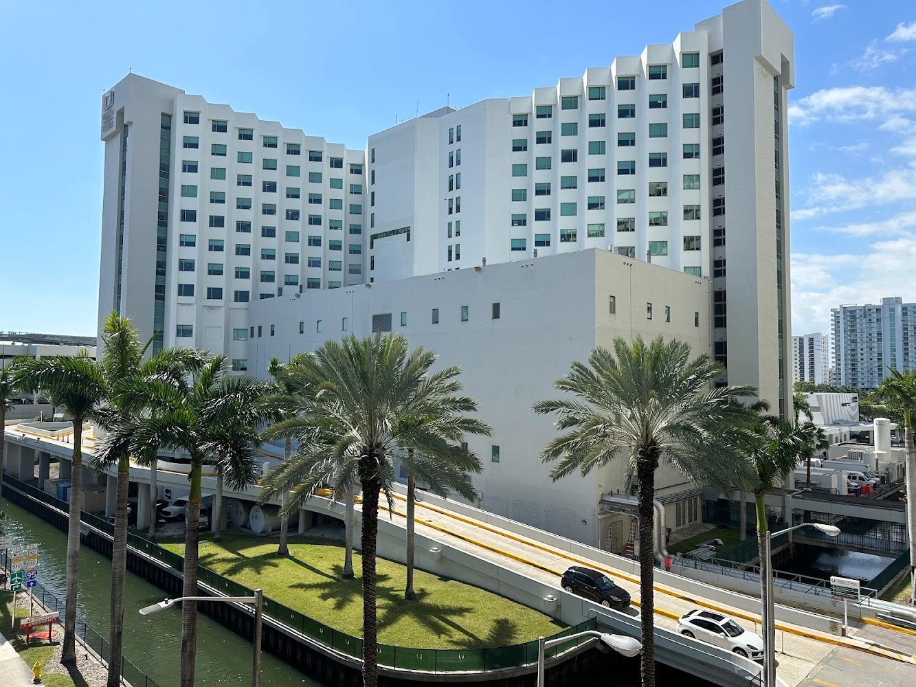
UHealth Tower

3.3

πŸ₯ Hospital

OpenΒ· Closes 5:00 PM Tue

At the University of Miami Health System (UHealth Tower Click to open side panel for more information and Jackson Memorial Hospital Click to open side panel for more information , orthopedic spine surgeonDr. Evan Trapana utilizes Ultra-Minimally Invasive Endoscopic Lumbar Foraminotomy. By using a camera about the size of a pencil, Dr. Trapana precisely opens the narrow nerve canal without cutting muscle or compromising spinal stability.

Understanding Neural Foraminal Stenosis and Lateral Recess Impingement

To understand why endoscopic foraminotomy is so effective, it helps to examine the anatomy of the nerve exit pathway:

[Main Spinal Canal] ──► [Lateral Recess Zone] ──► [Neural Foramen Exit Window] ──► Leg Nerves

β”‚ β”‚

β–Ό β–Ό

Hypertrophic Facet Joint Collapsed Disc / Bone Spurs

β”‚ β”‚

β””β”€β”€β”€β”€β”€β”€β”€β”€β”€β”€β”€β”€β”€β”¬β”€β”€β”€β”€β”€β”€β”€β”€β”€β”€β”€β”€β”€β”€β”€β”˜

β–Ό

Direct Pinched Nerve & Sciatica Pain

  • The Lateral Recess: The transitional zone inside the spinal canal just before the nerve enters the exit window. Hypertrophic (overgrown) facet joints or bony spurs here pinch the nerve early in its pathway.
  • The Neural Foramen: The exit tunnel itself. Loss of intervertebral disc height causes this window to collapse vertically, while disc bulges and thickened ligaments narrow it horizontally.
  • Double Impingement: Nerves can be compressed in both zones simultaneously, resulting in persistent radicular leg pain, numbness, and foot drop or weakness.

How Endoscopic Lumbar Foraminotomy Works

Unlike open back surgery, which approaches the spine through a long midline incision through back muscles, an endoscopic foraminotomy utilizes a transforaminal or interlaminar micro-corridor directly to the exit window.

[Micro-Incision (< 8mm)] ──► Dilates muscle fibers (Zero muscle cutting)

β”‚

β–Ό

[HD Micro-Endoscope Insertion] ──► Continuous fluid irrigation & 10x HD visualization

β”‚

β–Ό

[Targeted Bone & Ligament Removal] ──► Micro-burr widens exit window, freeing the nerve
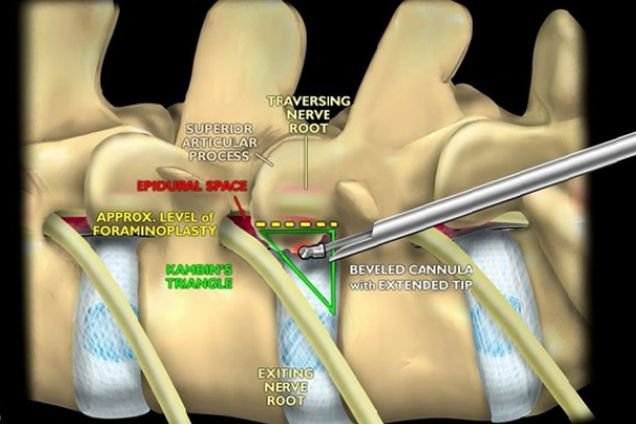
  1. Micro-Incision and Muscle-Sparing Dilation: Dr. Trapana creates a small micro-incision (less than 8 millimetersβ€”smaller than a dime). Specialized micro-dilators gently part muscle fibers along their natural planes without cutting them.
  2. HD Camera and Fluid Irrigation: A high-definition micro-endoscope is inserted into the foramen. Continuous sterile fluid irrigation washes away micro-bleeding and keeps the surgical field crystalline, giving Dr. Trapana a direct 10x magnified view of the compressed nerve root.
  3. Targeted Micro-Sculpting: Using diamond micro-burrs and microscopic punches, Dr. Trapana sculpts away only the precise bone spurs and thickened ligamentum flavum encroaching on the nerve, leaving healthy bone and joint structures untouched.
  4. Immediate Decompression: Once the nerve root is visibly decompressed and moving freely in its channel, the endoscope is removed, and a single band-aid or tiny suture closes the skin.

Endoscopic Lumbar Foraminotomy

Comparing Endoscopic Foraminotomy vs. Open Laminectomy/Fusion

Clinical Metric Traditional Open Surgery & Fusion Dr. Evan Trapana’s Endoscopic Foraminotomy
Incision Size 3 to 5+ inches Ultra-micro incision (< 8 mm)
Muscle Trauma Substantial muscle cutting & retraction Zero muscle cutting; gentle dilation
Bone & Joint Sparing Significant facet joint & lamina removal Selective bone sculpting; joint preserved
Spinal Stability High risk of instability (often forces fusion) 100% natural spinal stability preserved
Anesthesia Profile Full general anesthesia Light sedation / twilight or light general
Facility & Discharge Multi-day hospital stay Same-day outpatient discharge (1–2 hours post-op)
Recovery Window 6 to 12+ weeks Return to light daily activities in 3 to 7 days

Key Patient Benefits of the Endoscopic Approach

  • Preservation of Native Spinal Motion: By sparing the facet joints and major stabilizing ligaments, endoscopic foraminotomy eliminates the need for rigid screws, rods, or bone fusions.
  • Bypasses Prior Surgical Scarring: For patients who have had prior spine surgery, an endoscopic transforaminal approach reaches the exit nerve through clean, un-operated anatomical corridors, avoiding dense scar tissue beds.
  • Near-Zero Blood Loss & Low Infection Risk: The continuous fluid environment and micro-incision profile result in negligible blood loss and significantly lower infection rates compared to traditional open procedures.
  • Rapid Relief from Radiating Pain: Decompressing the exit nerve root directly at the source frequently provides immediate, post-operative relief from sharp shooting leg pain.

Who is a Candidate for Endoscopic Foraminotomy?

Endoscopic foraminotomy is an effective, targeted intervention for patients experiencing:

  • Severe Sciatica or Radicular Leg Pain caused by documented foraminal or lateral recess stenosis on MRI.
  • Numbness, Tingling, or Leg Weakness that has failed to improve with physical therapy, activity modification, or epidural steroid injections.
  • Recurrent Nerve Impingement following prior spine surgery where repeat open surgery carries high scar risks.
  • High-Risk Patients who want to avoid long general anesthesia times or complex inpatient hospital stays.

Conclusion

Persistent, shooting leg pain caused by a pinched exit nerve does not mean you have to undergo major open back surgery or spinal fusion. Through ultra-minimally invasive endoscopic lumbar foraminotomy, Dr. Evan Trapana provides patients across Miami and South Florida with precise, motion-preserving nerve relief and rapid recovery.

  • Practicing Specialist: Dr. Evan Trapana, MD (Orthopedic Spine Surgeon)
  • Academic Appointment: Assistant Professor of Orthopaedics, University of Miami Miller School of Medicine
  • Primary Practice Location: 1400 NW 12th Avenue, Suite 2, Miami, FL 33136
  • Clinical Affiliations: UHealth Tower Click to open side panel for more information & Jackson Memorial Hospital Click to open side panel for more information
  • Appointments & Consultations: 305-243-3000
logo